


Imaging and Diagnostic Methods
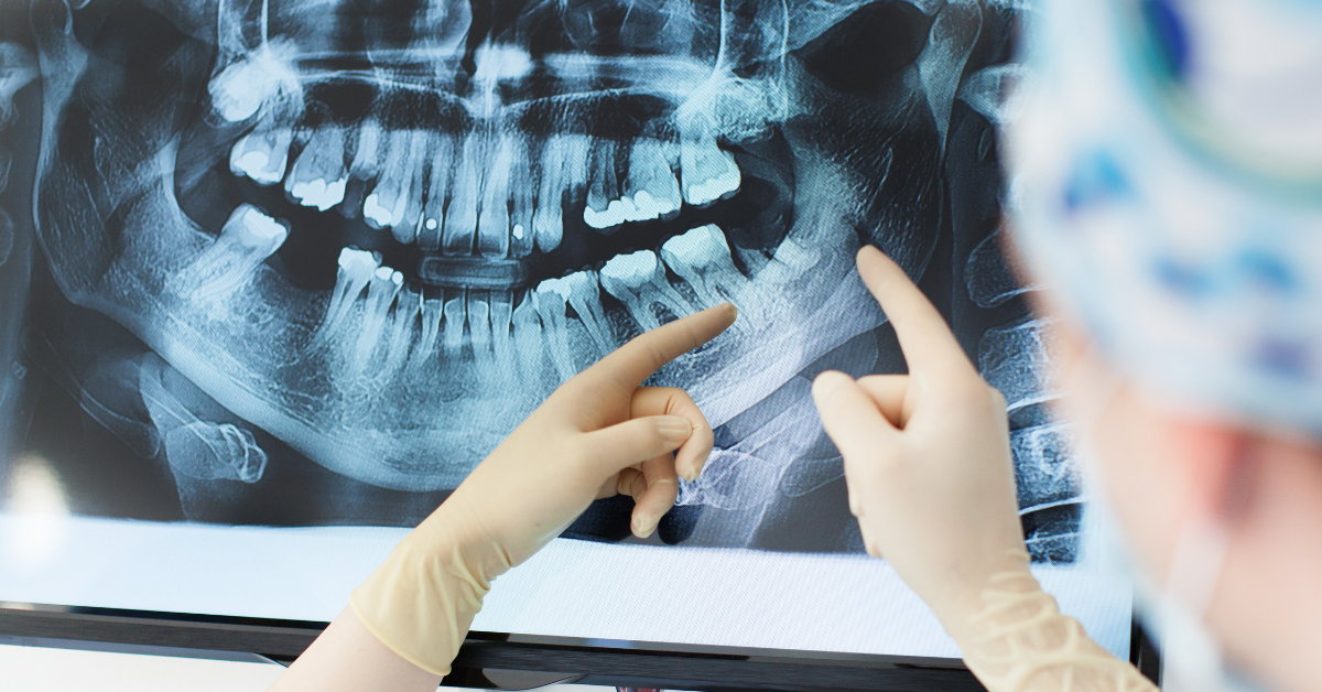
Imaging is an important tool in dentistry for treatment planning and diagnosis. Various radiographic methods are used to clearly see the condition of the teeth, gums, and surrounding tissues.
Panoramic X-Ray
It is a two-dimensional x-ray that provides a general view of the mouth, jaw, and teeth. It is used to detect all teeth, jaw bones, sinuses, and in some cases, temporomandibular joint disorders.
Cephalometric X-Ray
It is a profile x-ray of the head to evaluate the jaw structure and the relationship of the teeth to the face. It is used in orthodontic treatment planning to examine jaw and tooth development.
Periapical X-Ray
It is a more detailed imaging method that examines the root part of the tooth, the surrounding bone structures, and gum health. It is used to detect conditions such as root infections, cysts, and tumors.
Dental Tomography
Provides three-dimensional images of the teeth and jaw structures. It is ideal for detailed examination of the jaw bone structure when placing dental implants.
Hand-Wrist Radiography
It is an imaging method used especially in young patients before orthodontic treatment to evaluate jaw development and the placement position of the teeth. With this x-ray, we can estimate which stage of growth and development the patient is in. In orthodontics, we make decisions about the treatment of cases with skeletal problems based on this x-ray.
Bite-wing X-Ray
It is used to detect caries, tartar, and gum diseases by imaging the areas between the upper and lower teeth. It is especially important for the early diagnosis of caries.
Importance of Vitality Test
These are tests that evaluate whether the nerve function of the tooth is healthy. These tests are used to determine whether the tooth is alive, and therefore to determine an infection or the need for treatment.